
Whole Slide Imaging Best Practices: A Complete Guide to High-Quality Digital Pathology
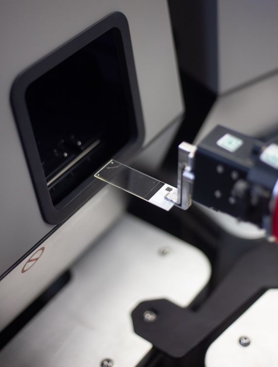
Digital pathology has fundamentally changed how researchers generate, analyze, and share histology data. Instead of relying solely on physical glass slides, laboratories can now digitize entire tissue sections into ultra-high-resolution images that are accessible from anywhere in the world. This technology, known as whole slide imaging (WSI), has become the foundation for modern pathology workflows, enabling everything from remote collaboration and quantitative image analysis to artificial intelligence (AI) development and large-scale digital archives.
As adoption continues to grow across academic research, pharmaceutical development, and clinical pathology, understanding how to produce high-quality whole slide images has become increasingly important. The quality of a digital slide directly impacts downstream image analysis, AI performance, data reproducibility, and publication outcomes.
In this guide, we'll explain what whole slide imaging is, how it works, why it has become essential for pathology research, and the best practices that help ensure consistently high-quality digital images.
What is Whole Slide Imaging?
Whole slide imaging (WSI), sometimes referred to as digital slide scanning or virtual microscopy, is the process of converting an entire glass microscope slide into a high-resolution digital image. Unlike traditional microscopy, where only one field of view can be examined at a time, whole slide imaging captures the entire tissue section in exceptional detail. Researchers can then zoom, pan, annotate, and analyze the specimen digitally, often at magnifications equivalent to 20x or 40x.
Today, WSI serves as the backbone of digital pathology. Researchers use whole slide images to collaborate across institutions, preserve valuable tissue specimens, develop AI algorithms, perform quantitative image analysis, and build searchable digital archives. Because digital slides can be shared instantly, whole slide imaging has dramatically reduced many of the logistical barriers associated with shipping fragile glass slides between laboratories.
As research continues to generate increasingly large datasets, whole slide imaging has become an essential tool for creating scalable, reproducible digital pathology workflows.
How Does Whole Slide Imaging Work?
Although scanning a microscope slide may appear straightforward, generating a high-quality whole slide image involves a carefully controlled workflow that begins long before the slide reaches the scanner.
Everything starts with proper tissue preparation. High-quality fixation, embedding, sectioning, staining, and coverslipping all play a critical role in determining the final image quality. Even the most advanced scanner cannot compensate for poorly prepared tissue or damaged slides.
Once slides are loaded into an automated scanner, sophisticated optical systems identify the tissue, establish precise focus, and capture thousands of overlapping high-resolution images across the entire specimen. Specialized software then stitches these individual images together into a seamless digital slide while correcting for differences in lighting, focus, and color consistency.
Before images are released, quality control is performed to identify common issues such as blurry regions, stitching artifacts, incomplete tissue capture, or focus inconsistencies. Once approved, the finished images are uploaded into digital pathology platforms where researchers can review, annotate, share, and analyze them remotely.
Because every stage of this workflow contributes to the final image quality, successful whole slide imaging depends on much more than simply owning a scanner.
Advantages of Whole Slide Imaging in Pathology
Whole slide imaging has become one of the most impactful innovations in pathology because it fundamentally changes how researchers interact with tissue samples. Digital slides eliminate many of the limitations associated with traditional glass slides while opening the door to entirely new analytical capabilities.
One of the greatest advantages is collaboration. Rather than shipping slides between institutions and waiting days for delivery, researchers can instantly share digital images with collaborators around the world. Multiple investigators can review the same slide simultaneously, making multidisciplinary studies significantly more efficient.
Whole slide imaging also improves data preservation. Physical slides naturally degrade over time and remain vulnerable to breakage or loss. Digitization creates permanent, high-resolution records that can be archived, searched, and revisited years later without risking damage to the original specimen.
Perhaps the biggest advancement has been the rise of quantitative digital pathology. Instead of relying solely on subjective visual interpretation, researchers can apply image analysis software to measure biomarker expression, immune cell infiltration, tumor burden, spatial relationships, and numerous other quantitative features. This improves reproducibility while reducing observer variability.
The growing adoption of artificial intelligence has further accelerated the importance of whole slide imaging. Machine learning algorithms require large collections of standardized, high-quality digital images to identify tissue patterns and generate reliable predictions. As AI becomes more integrated into pathology research, whole slide imaging provides the digital foundation these technologies require.
Best Practices for High-Quality Whole Slide Image Scanning
Producing high-quality digital slides begins long before a specimen enters the scanner. While modern scanning systems are highly automated, image quality is still heavily influenced by specimen preparation, scanner settings, and quality control procedures.
Proper tissue preparation remains the single most important factor. Well-fixed tissue, thin and uniform sections, consistent staining, and clean coverslips all contribute to better digital images. Small imperfections such as dust particles, fingerprints, mounting media residue, or tissue folds can interfere with autofocus and introduce artifacts that affect downstream analysis.
Selecting the appropriate scan resolution is equally important. While 40x scanning provides exceptional detail, many research applications can be adequately supported with 20x imaging, resulting in smaller file sizes and faster workflows. Choosing the appropriate magnification based on project objectives helps balance image quality, storage requirements, and scanning efficiency.
Consistency should also be a priority throughout large studies. Standardizing scan settings, color calibration, compression methods, and file naming conventions improves reproducibility and creates datasets that are better suited for image analysis and AI development.
Finally, every digital pathology workflow should include rigorous quality control before images are delivered. Reviewing scans for focus issues, stitching artifacts, incomplete tissue capture, and other common defects helps prevent costly rescanning later in the research process.
Challenges & Limitations of Whole Slide Imaging
Despite its many advantages, implementing whole slide imaging at scale presents several operational challenges. Digital pathology programs require specialized equipment, robust data storage, technical expertise, and standardized workflows to consistently produce high-quality images.
One of the biggest obstacles is data management. Whole slide images are often several gigabytes in size, making storage, sharing, and long-term organization increasingly difficult as projects grow. Without an efficient digital pathology platform, managing thousands of large image files can quickly become overwhelming.
Image quality is another common challenge. Variability in tissue preparation, staining quality, scanner calibration, or autofocus performance can introduce inconsistencies that reduce the reliability of image analysis and AI applications. Maintaining standardized workflows across large studies requires both experienced personnel and rigorous quality assurance.
Scaling digitization projects also presents logistical hurdles. Many research organizations lack the infrastructure needed to scan tens of thousands of slides while maintaining rapid turnaround times and consistent quality. Purchasing scanners, maintaining equipment, training staff, and managing digital infrastructure can represent a significant investment.
HistoWiz was built to remove these barriers. Rather than simply providing slide scanning, HistoWiz offers a complete digital pathology workflow designed to support researchers from sample preparation through digital analysis. High-throughput scanning capabilities allow laboratories to efficiently digitize everything from small research studies to large retrospective archives while maintaining rigorous quality control throughout the process.
Once scanned, images can be securely managed within PathologyMap™, HistoWiz's cloud-based digital pathology platform, where researchers can organize, annotate, review, and share slides without the burden of maintaining their own storage infrastructure. For teams looking to move beyond image acquisition, HistoWiz also provides advanced image analysis services, helping researchers transform high-quality digital slides into meaningful quantitative data.
By combining automated scanning, standardized workflows, cloud-based image management, and scientific expertise, HistoWiz enables research teams to adopt digital pathology without the complexity of building and maintaining their own scanning infrastructure.
Accelerate Your Digital Pathology Workflow with HistoWiz
Whole slide imaging has become an essential component of modern pathology research, enabling faster collaboration, improved data preservation, quantitative image analysis, and AI-driven discovery. However, generating consistently high-quality digital slides requires more than advanced scanning hardware—it requires standardized workflows, experienced quality control, and an integrated digital pathology platform.
HistoWiz provides end-to-end whole slide imaging services designed to help researchers digitize slides with confidence. Whether you're scanning a small validation study or building a large digital pathology archive, our automated workflows, expert quality assurance, and cloud-based PathologyMap™ platform make it easier to move from glass slides to actionable scientific insights.