
PathologyMap Platform Updates: Fall 2026
In This Article
Over the past three months, we shipped a steady stream of improvements to PathologyMap, the HistoWiz platform for viewing, sharing, and analyzing your histology data, all with one goal: less waiting, more science. Nearly everything below started as feedback from researchers like you. Here's a roundup of what's new from August through early November 2026.
Compare Slides in Perfect Sync
Turn on the new Sync Controls and every slide pans and zooms together. Line up an H&E against its IHC stains, or treated against control, and compare them region by region. A toggle right above the slides switches syncing on or off, so you can line slides up first and then explore them together.
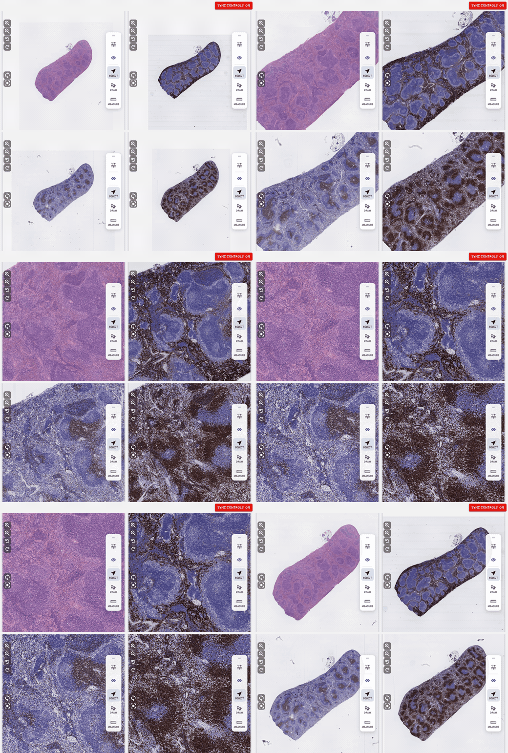
H&E, F4/80, CD3 and B220 stains of one mouse spleen section, locked together.
The brightfield viewer itself also runs on an upgraded imaging engine, so slides open faster and navigation is smoother.
See Exactly What the Image Analysis Found
Image analysis orders now come with more than a report and data. You also get annotated slides: every cell the analysis detected and classified, drawn right onto the slide. Open an annotated slide next to the original, turn on Sync Controls, and check the results region by region, all the way down to single cells.
Contact your sales rep to set up Image Analysis with HALO on your next order.
Draw, Measure and Download Your Annotations
The brightfield viewer has a new annotation rail beside the slide. Pick a tool, pick a color, and mark it up.
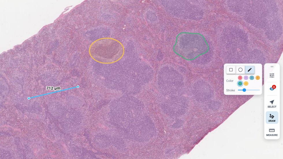
The draw tools beside a mouse spleen H&E: an ellipse, a pencil outline and a saved 712 µm ruler measurement.
- New ellipse and pencil tools. Every shape shows up in the color you picked, straight away, and pressing Esc cancels whatever you're in the middle of.
- Measurements that stick. Ruler measurements save as annotations, stay put as you zoom and scroll, and reload with the slide. Select one to drag either end, or delete it with the Delete key or the trash icon.
- Take them with you. Download Annotations sits right under Download Slide and exports your annotations in a format compatible with HALO and ImageScope.
Your IF Viewer, Now 3x Faster
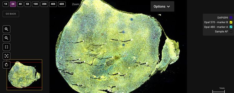
We rebuilt the engine behind our immunofluorescence viewer. Zooming in and out is now about 3x faster than before, so you can dive from the whole slide down to single cells and back without missing a beat.
- 15 channels at once, up from 6. Toggle any marker on or off and keep your entire multiplex panel in view.
- Glide through huge scans. Pan and zoom stay smooth while image tiles stream in, and slides open faster than ever.
- 40x zoom in one click, and up to 60x when you need it.
- More image, less clutter. Panels tuck away when a slide opens, full slide names are always visible, the zoom-limit pop-ups are gone, and the controls are larger and higher in contrast, with tooltips.
Turn IF Images to Any Angle
Click the new Rotate button in the IF viewer and a dial opens over the slide. Drag it to line the tissue up exactly the way you want (hold Shift to snap to 15°), or type in an exact angle. Shared links reopen at the same spot and angle, and the brightfield viewer has the same dial.

The rotation dial over a 7-plex IF slide, turned to 35°.
Share an Entire IF Order in One Click
Open the new gear menu in the IF viewer, turn on Share IF Order, and send the link. Collaborators get every IF slide in the order, in a viewer and a slide grid, so your PI, your pathologist and your bench team are all looking at the same thing. No file shuffling required.

Gear menu → Share IF Order: On → Copy link.
Want an expert read on your multiplex IF? Contact us to request an IF pathology consultation.
All Your Pathology Consultations in One Place
The new Consultations page in the sidebar lists every pathology consultation for your team, with its status and due date at a glance.

The Consultations page, with a completed consultation's details and progress.
- Open one to see everything: its slides, annotations and request details.
- Read the final report right on the page. The report PDF opens inline, with any attached files listed underneath.
- Find it fast. Consultations, and image analysis reports, now show up in the search bar too, with the matching text highlighted.
Your Data, Your Way
- Block Manager → Excel in one click.
- Group slides by order in the slide grid.
- Snappier dashboards for Team Overview, the newsfeed and slide groups.
- Team Overview shows pathology consultation and image analysis report columns on every orders tab.
Smoother Ordering
- Better sorting when picking slide services.
- Locked orders stay locked. Once an order is locked for processing, its order form can no longer be changed by mistake, and a notice explains why.
Uploading Your Own Scans
If you upload your own whole slide images, PathologyMap now converts Akoya .qptiff multiplex files and handles Zeiss .czi files as brightfield slides. Uploaded slides appear in your account only after conversion finishes, so you never open a slide that is still processing.
Automatic Label Scrubbing for Sensitive Samples
For projects that need it, slides can be automatically scrubbed of identifying information when they are received. Label images, macro images, thumbnails, and file metadata are removed. A neutral label with the slide's unique ID takes their place, so every slide can still be traced. Scrubbing covers SVS, OME-TIFF, .qptiff, and DICOM files. Contact us if you'd like it turned on for your account.
Explore Our IHC Antibody Catalog
Our validated IHC antibody catalog has a brand-new home. Browse the full list or filter by application, species or manufacturer. Every antibody has its own page, ready to bookmark and share.
Sneak Peek: Shipping Manifests
We can now create shipping manifests for the samples we ship back to you: every block and slide in the box, listed and ready to check off when it arrives.

A sample manifest (example data).
Want one in your next return box? Just ask your customer service rep, listed on your Team Overview page.
And a Few More Good Things
- A fresh, simpler sign-in page.
- Fewer pop-ups between you and your slides.
It's All Live Now
Nothing to install, nothing to update. Log in to PathologyMap to try it out. Got an idea for what we should build next? Use Request Feature in the PathologyMap sidebar. We read every single one, and we have more big things in the works.